
:quality(85)/cloudfront-us-east-1.images.arcpublishing.com/infobae/76Q3V4IS6W7CAP5TT6MVJGCHMQ.jpg)


:format(jpeg):focal(2425x455:2435x445)/cloudfront-us-east-1.images.arcpublishing.com/gfrmedia/VHMDSLP56BEZDI2PZFVOPDHJGA.JPG)

to: Dr. Mario Osorio ValeroAnd Head of the TC District of the ISSSTE National Medical Center “20 November”.
LaSalud.mx.- Computed tomography (CT) is considered One of the most important inventions in medicine in the twentieth centuryFor being undoubtedly the most important event that occurred in the field of radiological diagnosis, after the discovery of X-rays in 1895. Tomography is the first imaging technology where digital procedures can be applied from the first device manufactured.
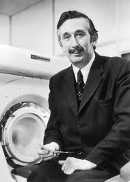
Since the Austrian mathematician J. radon, who showed in 1917 that it was possible to reconstruct an image of a two- or three-dimensional object from a large number of its projections, passing through Alan MacLeod Cormac, who proved in 1963 that it is possible to obtain a cross-sectional image by solving multiple mathematical equations, even Godfrey Newbold Hounsfield who managed to create the first tomography prototype; The necessary link between scientific and technical development in all branches of science and technological innovation has been highlighted to enable the creation of tomography with its indisputable diagnostic efficacy. Cormack and Hounsfield got The Nobel Prize in Physiology and Medicine in 1979. It was a very important event The first time the Nobel Prize in Medicine was awarded to two non-medical scientists.
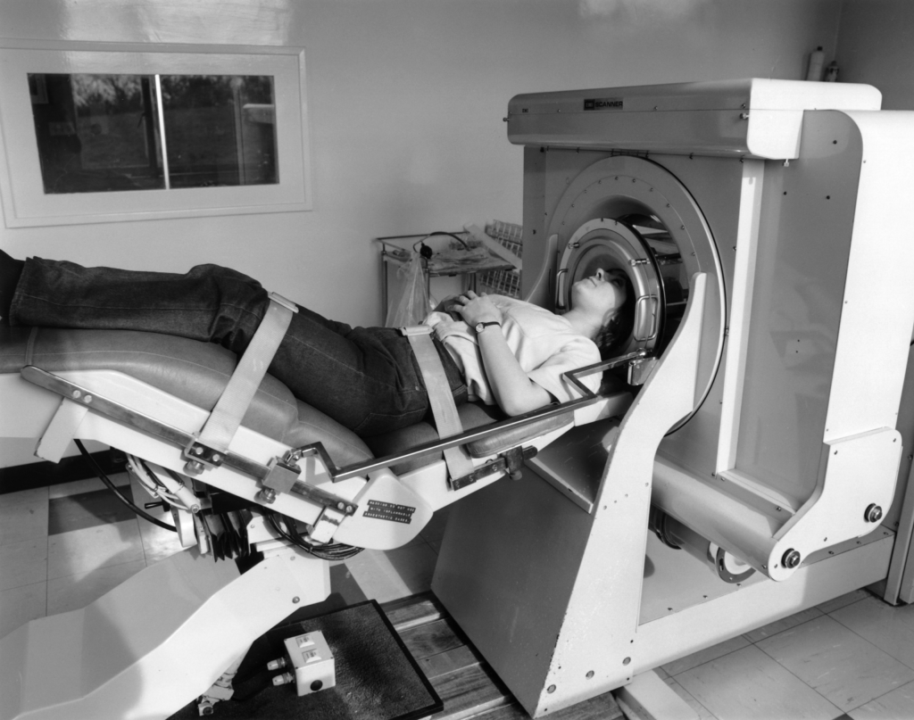
The history of tomography is taking its first steps October 1971, the year the first CT scan was performed on a 41-year-old patient To suspect a tumor in the frontal lobe using a prototype device in Atkinson Morley Hospitalfrom London; the I’m Mark I, Built by Godfrey Hounsfield and his team at EMI (Electrical Music Industries, Ltd) Central Research Laboratories Corporation, East London.
With this study, the patient’s diagnosis was confirmed and the operation was successfully performed Surgeon’s wordsThe tumor was just like the picture“. Here began the contribution of the technological revolution to the health of mankind.
The first commercial tomography was introduced in 1973, using a linear beam with a single detector and translation/rotation mechanism, had an 80 × 80 matrix—we currently work with 160 × 160 to 512 × 512 matrices with a spatial resolution of 0.5 cm—and required the use of a water bag for image stabilization and normalization. Each image needs 4.5 minutes to acquire and an additional 20 minutes to rebuild, so At first it was only used to visualize immobile parts of the body such as the skull.
Over the next decade and a half, significant technical improvements were made that reduced acquisition time, allowing exploration of the entire body, as well as the skull. This development opened the possibility of obtaining images of the whole body and allowed the diagnosis of lesions not only of the central nervous system, but also the rest of the diseased organs.
Computed tomography has progressed in recent years towards obtaining volumetric images of a specific body segment, which are obtained while sliding the table in which the patient is located, mainly due to the increase in detectors, with which multiple segments” are achieved in a single helical scan to increase spatial accuracy.
in a 1989, helical tomography appeared, which had significant advantages over its predecessor, computed tomography (CT). Because it allowed the continuous acquisition of several images for each inspiration; This was possible due to the synchronization between the x-ray tube, the table and the detectors (one row of detectors).
in the year 1998, multi-detector tomography was born, also known as Multislice (TCMS), this technology has 4 rows of detectors that increased with the mastery of instrument design and technology until today we find tomography with 512 rows of detectors, it is important to emphasize that the more rows of detectors the better the results. In the case of tomography with 512 rows of detectors, the technology is smart heartTo explore the heart with images of the coronary arteries without movement and at any heart rate. its technology Intelligent Smart Dose Reduces radiation exposure by up to 82%; On the other hand, technology smart strike It allows critical decisions to be made regarding the condition of patients with multistage cerebral perfusion protocols.
Technological advances allow this imaging method to be applied in different fields of medicine with its own benefits Making diagnoses that up to 50 years ago were impossibleAn example is in the field of pediatrics, where we can perform studies without anesthesia with a complete exploration of the chest and abdomen in less than a second.
The continuous development of computed tomography has led to the modification of many health-related activities over time, due to the astonishing development of technology. There are currently a few medical procedures that do not involve the application of this diagnostic imaging modality.It is the product of a precipitous development of science and technology that is modifying the way knowledge is conceived and applied in medical activities and health services around the world.
This, and other interesting articles, are accompanied by reports; Interviews and private collaborations with the most famous local and international specialists; You can find it in our next special multimedia issue of LaSalud.mx: “Imaging, Radiation Oncology, Nuclear Medicine” with an Ibero – Latin American presence.

“Social media evangelist. Student. Reader. Troublemaker. Typical introvert.”
More Stories
Medicine practiced by men versus medicine practiced by women
The College of Humanities, Social and Educational Sciences celebrates the graduation of 397 male and female students
Japanese ancestral ritual that is an effective and quick cleaning method: in 15 minutes organize your home and help emotional well-being